Radiological diagnostics
Digital radiography is the achievement of technology without which it is certainly hard to imagine a modern dental office. Radiological study is the basis of the oral examination at the first dental visit, because it allows non-invasive assessment of the health of your teeth and surrounding tissues, especially those that are invisible to the naked eye. This test also enables a dentist to prepare a personalized treatment plan that takes into account individual differences in the oral structure of each patient.
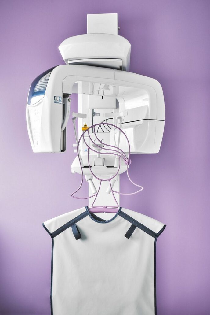
Thanks to the use of modern equipment, we can ensure fast and safe production of high quality radiological images. The photos are displayed on the computer screen immediately after they are taken, where they can be further digitally processed in order to reveal additional details that are diagnostically relevant.
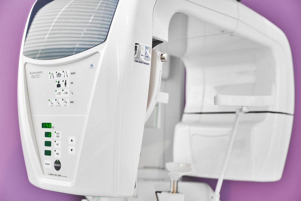
Radiological examination is safe for the patient provided that the principles of safety and radiological protection are applied, consisting in using the lowest possible radiation dose adjusted to the patient’s weight, as well as special lead aprons protecting the patient’s trunk and neck against radiation.
Paediatric radiology
Digital radiography is completely safe for a child due to the use of a reduced dose of radiation and paying special attention to the radiological protection of a small patient during the examination. When it comes to children, radiological tests are extremely helpful in routine assessment of the health of milk teeth, as well as in determining any abnormalities in permanent tooth structure and placement.
According to the valid regulations, children under 16 who are going to be performed radiological examination have to bring the child’s health book with them, in which the dentist notes its performance.
Radiological examination in pregnancy
As far as pregnant women are concerned, radiological diagnostics is not performed, except for emergency clinical cases.
To ensure protection of the mother and child against X-rays, it is extremely important to inform your dentist about your pregnancy or its suspicion during your first visit.
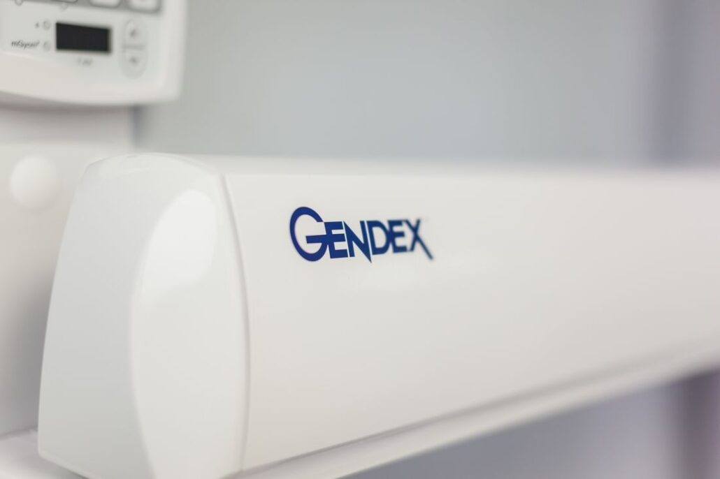
In our office, we use only modern equipment that guarantees patient’s safety and comfort:
• Gendex 765DC X-ray system with VISUALIX digital radiovisiography system – for intraoral photos
• Gendez GXIO-770 X-ray with GXS-700 digital radiovisiography system – for intraoral images, which thanks to the latest technologies allows a significant reduction of the radiation dose while maintaining the high resolution of created images.
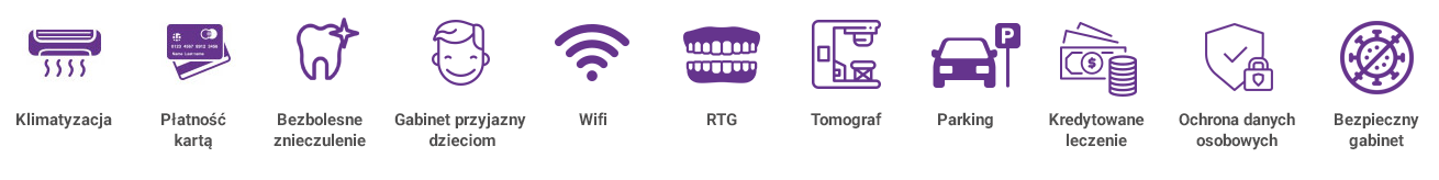
• Morita Veraviewepocs 2D camera – for pantomographic images of the Japanese company called Morita, which stands out among its competitors with extraordinary innovation and precision. That is why, the equipment offered by the company makes it possible to shorten the exposure time and limit the dose of absorbed radiation to one of the lowest on the market thus maintaining the excellent quality of the photo taken.
Types of tests performed
Pantomographic (panoramic) photo
It reveals the whole of both dental arches, jaw and mandible bones, maxillary sinuses and temporomandibular joints. It is invaluable for accurate diagnosis and verification of the previously performed treatment. It is mainly used for the basic assessment of the dentition, as well as for planning procedures in the field of dental surgery, implantology and orthodontics.
Intraoral image (point)
It is a photo of a single tooth or a few neighboring ones which when enlarged shows particular tooth structures and also periapical tissues near the roots of the teeth. In endodontics the photo is necessary to control the course of root canal treatment, while in conservative dentistry it enables to identify carious defects accurately and to determine if the previously apllied fillings are tight enough.
Cephalometric image
It depicts hard tissues and the outline of soft tissues within the patient’s bony face. Thanks to it, it is possible to diagnose malocclusion and to make detailed measurements which will be necessary in orthodontic treatment.
CBCT computed tomography
This study shows the tissues of facial skeleton and allows them to be imaged in 3D. This enables to assess the facial bone structure accurately, which is necessary to plan and carry out implant treatment. It is also used by other areas of dentistry, such as endodontics, orthodontics and dental surgery.
Cephalometric photos and computed tomography are possible to be done at RTG DENT Dental Radiography which our office has established cooperation with.